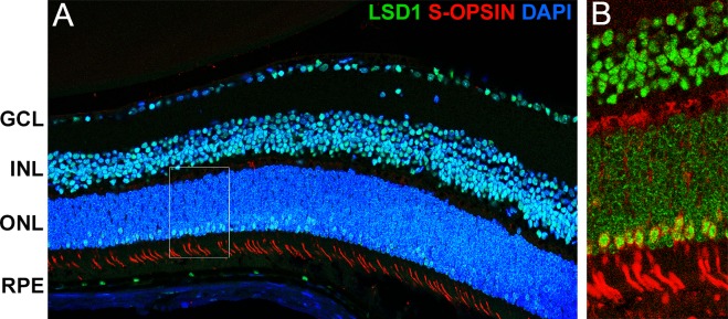
Figure 5.
High levels of LSD1 in cone photoreceptors but not rods. Immunofluorescence staining of P36 C57BL/6J mouse retinas for LSD1 (green), short wavelength cone opsin (red), and DAPI nuclear stain (blue). The 40× merged image taken with a confocal microscope showed LSD1 expression in all three nuclear layers and short wavelength cone opsin expression in cone photoreceptor outer segments (A). The 60× image shows perfect correlation between cells with high LSD1 expression (green) along the outer edge of the ONL and the cone opsin (red) (B).

