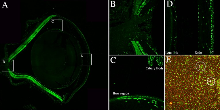
Figure 9.
LSD1 is expressed throughout the murine eye. Immunofluorescence of LSD1 (green) and DAPI (blue) in P36 C57BL/6J retinal sections and RPE flatmount (A–E). The 20× confocal image of an entire P36 murine eye stained with LSD1 (green) (A) showed LSD1 expression throughout many ocular structures. White boxes indicate areas from which 40× images were taken for B–D to focus on various ocular structures outside of the retina including the optic nerve (B), lens (C), and cornea (D). In C the bow region of the lens and ciliary body are labeled, and in D the lens, iris, corneal endothelium, and corneal epithelium are labeled. A 40× image from an RPE flatmount showed LSD1 nuclear expression in green and ZO-1 in red (E) to outline all RPE cell borders. Two white circles with arrows highlight different RPE cells that contain either one or two nuclei.

