Fig. 2.
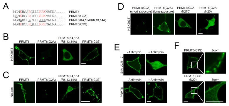
Characterization of the PRMT8 plasma membrane-targeting domains. (A) Schematic diagrams of point mutations within the N-terminal 20 amino acids of PRMT8. Mutated amino acids are underlined. Basic amino acids are colored in red. (B, C) Cellular localization of PRMT8-GFP, PRMT8(G2A)-GFP, and PRMT8(K4,15A/R6,13,14A)-GFP in HEK293T cells (B) and in cultured cortical neurons (C) Scale bar, 20 μm. (D) Cellular localization of PRMT8(G2A)-GFP, PRMT8(G2A/N270)-GFP, and PRMT8(G2A/N20)-GFP in HEK293T cells. Scale bar, 20 μm. (E) Effects of phosphoinositide depletion on the plasma membrane localization of PRMT8-GFP and Aplysia PDE4 short-form, S(N-UCR1/2)-GFP after antimycin treatment. Images were acquired before and after treatment with 10 μM antimycin for 40 min. Scale bar, 20 μm. (F) Cellular localization of PRMT8(C9S)-GFP and PRMT8 (C9S/N20)-GFP in HEK293T cells. Scale bar, 20 μm.

