
Figure 13.
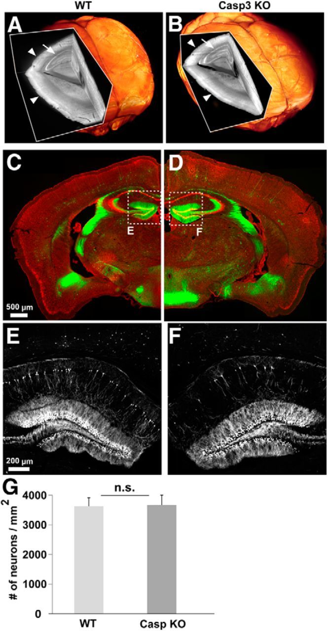
Anatomical characterization of caspase-3 KO brain. 3D scans of whole brains from 10-month-old wild-type (left) and caspase-3 KO (right) mice crossed to GFP-M line. The brains were cleared and subsequently imaged using light-sheet ultramicroscopy (Ertürk et al., 2012b). There was no gross abnormality of capase-3 KO brains compared with control mice: the cortex (B, arrowheads) and hippocampus (B, arrow) of the caspase-3 KO mice was similar to wild-type mice (A). C–D, Coronal brain sections from 3-month-old caspase-3 KO (right) and wild-type littermate (left) mice. Sparse neurons are labeled with GFP (green) and neuronal cell bodies are labeled with Nissl staining (red). No gross anatomical abnormality was observed in caspase-3 KO brain (see text for details). E, F, Hippocampus of wild-type (E) and caspase-3 KO (F) mice; only GFP images are shown. G, Quantification of Nissl staining in the cerebral cortex showing no difference in the numbers of neurons per square millimeter between wild-type and caspase-3 KO mice (p = 0.9311 calculated by Student's t test). n = 11 cortex scans for wild-type and 6 for caspase-3 KO from at least 3 different mice per group.