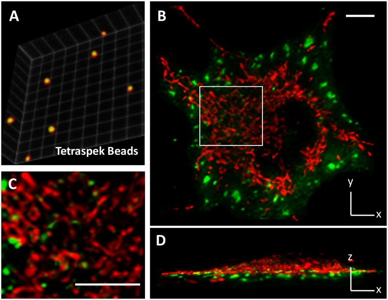
Figure 2. Dual-color LLSM imaging of metabolic condensates in live cells.
(A) Dual color calibration with TetraSpek™ microspheres (200 nm in diameter) that are embedded in polyacrylamide gel. The centroids of red and green images are overlapped with subvoxel precision. Calibration factors are applied to dual-color data. (B, C and D) A representative image shows the spatial distribution of glucosomes (green, hPFKL-mEGFP) and mitochondria (red, MitoTracker Red CMXRos, Cat# M7512) in a Hs578T cell. The indicated region of interest (a white box) is enlarged for clarification. Scale bars, 10 um.

