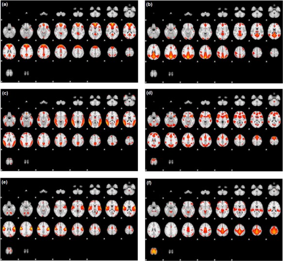
Figure 3.

Temporal lobe epilepsy dataset: Z‐score maps of components of interest, overlaid in Montreal Neurological Institute (MNI) space: (a) Anterior DMN, (b) Posterior DMN, (c) Alerting network, (d) Salience network, (e) Premotor cortex, and (f) Primary somatosensory cortex. Orientation is radiological. [Color figure can be viewed at http://wileyonlinelibrary.com]
