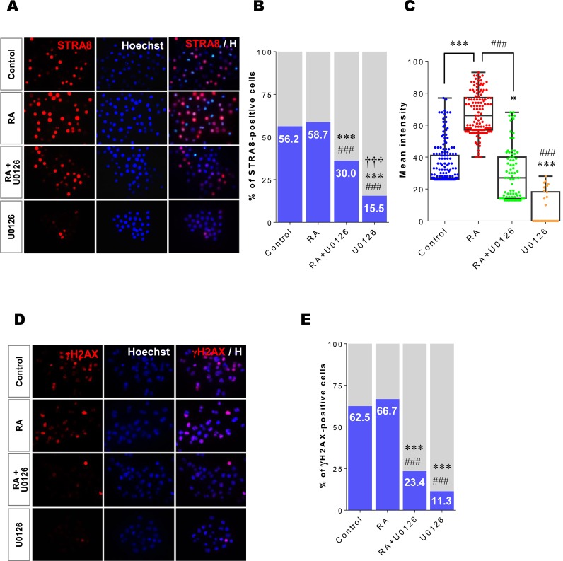
Fig 4. Inhibiting ERK1/2 activity suppresses STRA8 protein expression and meiotic initiation in XX germ cells.
Isolated XX germ cells at E12.5 were cultured under four different conditions (control, RA, RA+U0126, U0126) for 24h. (A) After culture, germ cells were immunostained with an anti-STRA8 antibody (red) and counterstained with Hoechst33342 (blue). (B) The ratio (%) of STRA8-positive cells in each culture condition was estimated using ImageJ software. *** p < 0.001 vs. control; ### p < 0.001 vs. RA; ††† p < 0.001 vs. RA+U0126. (C) The fluorescent intensity of STRA8 protein expression in each positive cell was quantified using ImageJ software. The results were represented as a box-whisker plot. Each dot represents a single analyzed germ cell. Each box indicates the middle 50% of the data and midline in the box indicates the median. The whiskers extend to the most extreme data point, which is no more than 1.5 times the quartile range. * p < 0.05, *** p < 0.001 vs. control; ### p < 0.001 vs. RA. (D) To examine the entry into meiosis, cultured XX germ cells were stained with an antibody against γH2AX (red) and counterstained with Hoechst33342 (blue). (E) The ratio (%) of γH2AX-positive cells in each condition was estimated using ImageJ software. *** p < 0.001 vs. control; ### p < 0.001 vs. RA.

