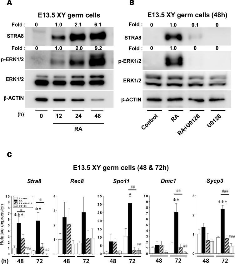
Fig 5. The effect of RA-stimulated ERK1/2 activity on the meiotic initiation in XY germ cells.
(A) Isolated XY germ cells at E13.5 were cultured with 1μM RA for 0, 12, 24, and 48h, and the ERK1/2 phosphorylation and STRA8 protein expression were then analyzed using Western blotting. (B) Isolated XY germ cells at E13.5 were cultured under four different conditions (control, RA, RA+U0126, U0126). After 48h culture, the cells were subjected to Western blotting to quantify the ERK1/2 phosphorylation and STRA8 protein expression levels. The fold changes of these proteins were represented on the top of each as numerical values that calculated relative to either 0h level (A) or the control (B) set as 1.0. (C) Isolated E13.5 XY germ cells were cultured under four different conditions (control, RA, RA+U0126, U0126) for 48 and 72h. After culture, the cells were subjected to qPCR analysis to determine the transcript levels of Stra8 and meiotic marker genes (Rec8, Spo11, Dmc1 and Sycp3). The expression levels were normalized to β-actin mRNA expression. All expression values were calculated relative to control levels set at 1.0. Data represent the mean ± SEM (n = 3–6). * p < 0.05, ** p < 0.01, *** p < 0.001 vs. control; # p < 0.05, ## p < 0.01, ### p < 0.001 vs. RA.

