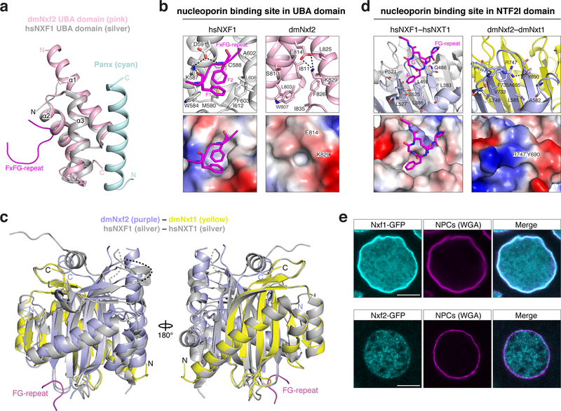
Figure 6. Nxf2 lost nucleoporin binding.
a, Superposition of the overall structure of dmNxf2 UBA domain (pink) complexed with Panoramix helix (cyan) and the crystal structure of hsNXF1 UBA domain (PDB ID: 1OAI) (in silver) 35.
b, Left: Ribbon (top) and electrostatic surface (bottom) representation showing the FG-repeat binding pocket in the crystal structure of hsNXF1 UBA domain (gray) in complex with a nucleoporin FxFG-repeat peptide (magenta; PDB ID: 1OAI) 35. Right: Ribbon (top) and electrostatic surface (bottom) representation showing the closed putative FG-repeat binding pocket in the crystal structure of dmNxf2’s UBA domain (pink).
c, Superposition of the overall structure of dmNxf2’s NTF2-like domain complexed with dmNxt1 (purple and yellow) and the crystal structure (silver) of hsNXF1–NXT1 (PDB ID: 1JKG) 39 (dashed curves: invisible loops in the structure).
d, Left: Ribbon (top) and electrostatic surface (bottom) representation showing the specific recognition of nucleoporin FG-repeat (magenta) in the hsNXF1 (light blue)–NXT1 (gray) structure (PDB ID: 1JN5) by a hydrophobic FG-repeat binding pocket 39. Right: Ribbon (top) and electrostatic surface (bottom) representation showing the blocked putative FG-repeat binding pocket in the crystal structure of Nxf2’s NTF2-like domain (purple) complexed with Nxt1 (yellow). The hydrogen bond is shown as black dashed line.
e, Confocal images showing individual nurse cell nuclei from flies expressing GFP-Nxf1 (upper row) or GFP-Nxf2 (lower row) (scale bar: 5 μm). Nuclear pore complexes (NPCs) are stained with wheat germ agglutinin (WGA).

