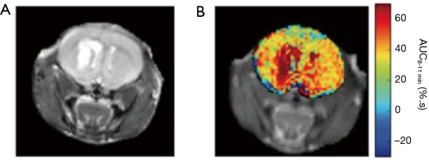
Figure 6.
3-O-Methyl-D-glucose (3OMG) chemical exchange saturation transfer (CEST) MRI of malignant brain tumor. (A) Anatomical image of the mouse brain. (B) The area under the curve image calculated for the last three minutes of the CEST scan (using a single CW magnetization transfer prepulse of strength 1.5 µT and duration 2 s), a period of 8–11 min post injection of 3OMG (3 g/kg, 1.9M, 200 mL). Figure taken with permission from (59).

