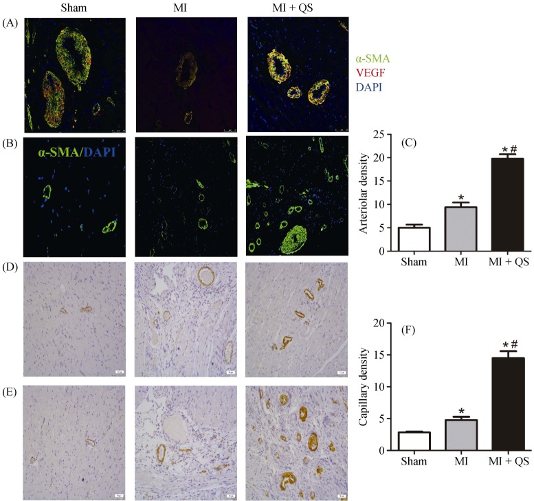
Figure 5. Immunohistochemistry and immunofluorescence assessment of myocardial angiogenesis.
(A): Immunofluorescence for the expression of angiogenesis positive for VEGF and α-SMA in the vascular endothelium; (B): sections were stained with α-SMA staining for the arteriolar density measurement; (C): quantification of the arteriolar density; (D): CD31 immunostaining to identify capillaries in the peri-infarct region; (E): CD31 immunostaining to identify capillaries in the infarct region; and (F): quantification of the capillary density. n = 6 in per group. The magnification for A is 400 ×, for B, D and E is 200 ×. *P < 0.05 compared with the Sham group, #P < 0.05 compared with the MI group. MI: myocardial infarction; QS: Qishen capsule.

