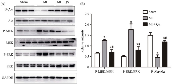
Figure 7. The activities of the Akt and MEK/ERK1/2 pathways were detected with western blotting.
(A): The phosphorylation of MEK and ERK1/2 was attenuated markedly by QS in the post-infarct hearts, whereas the phosphorylation of Akt increased; and (B): the histogram shows the relative protein levels of the phosphorylation of Akt and MEK/ERK1/2. The data represent the results of three separate experiments. The results are expressed as the means ± SD. *P < 0.05 compared with the Sham group, #P < 0.05 compared with the MI group. Akt: protein kinase B; ERK: extracellular regulated protein kinase; GAPDH: glyceraldehyde-3-phosphate dehydrogenase; MEK: mitogen-activated protein kinase; MI: myocardial infarction; QS: Qishen capsule.

