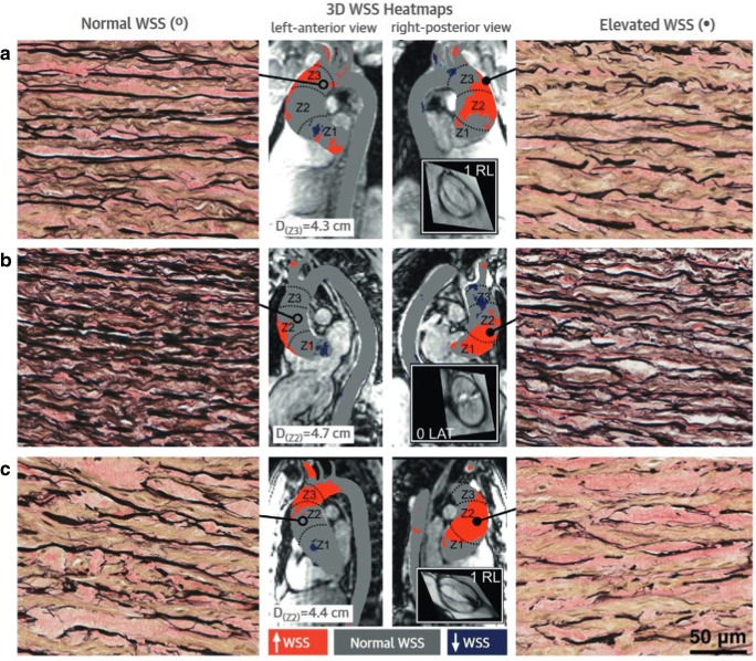
Fig. 5.
Aortic wall specimens of regions with normal WSS (left panels) and high WSS (right panels) in three (a–c) patients with BAV aortopathy (× 40 magnification). Note the decreased number of elastin fibers (black) in the context of elevated WSS. Center panel: 4D flow MRI–based maps depicting areas with increased (red) and depressed (blue) WSS. Reprinted from Guzzardi et al, Valve-related hemodynamics mediate human bicuspid aortopahy, J Am Coll Cardiol 2015;66:892–900, with permission from Elsevier

