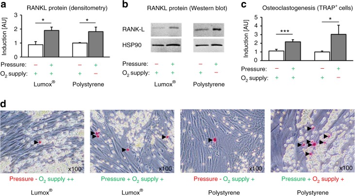
Fig. 5.
Effects of mechanotransduction vs. oxygen supply on hPDL-fibroblast-mediated osteoclastogenesis. a Densitometric immunoblot analysis of membrane-bound RANKL protein expression (N = 5). b Representative immunoblot of membrane-bound RANKL protein expression. c Quantification of TRAP-positive osteoclast-like cells per coculture well (N = 3, n = 9). d Representative images (×100) of coculture TRAP staining. TRAP-positive cells appear red (black arrows), RAW264.7 spherical osteoclast-precursor cells appear yellow and spindle-shaped hPDL fibroblasts are transparent. Bars indicate mean values ± standard deviation. *P ≤ 0.05, **P ≤ 0.01, ***P ≤ 0.001. AU = arbitrary units

