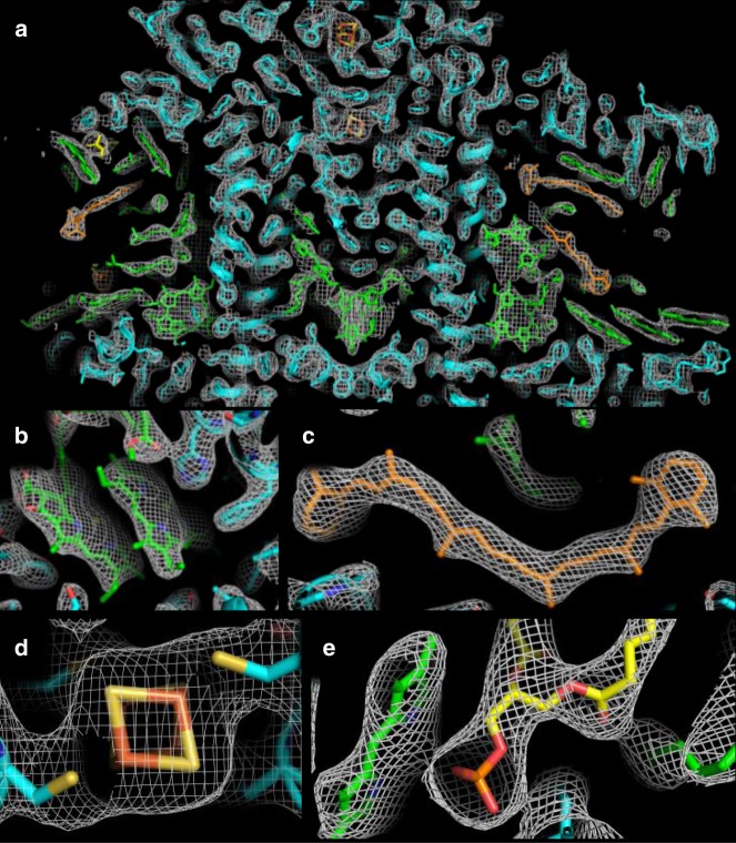
Fig. 3.
Electron density map (2Fo–Fc at 1.5σ) and model of various PSI structural elements of the XFEL structure of PSI. In all images, protein is colored cyan, chlorophyll (Chl) molecules are colored green, β-carotenes are colored orange, and lipids are colored yellow. In panels b–e, nitrogen atoms are colored blue, oxygen atoms are colored red, and magnesium atoms are colored bright green. a A slice through the center of electron density of a monomer of PSI is shown, b the electron density of the “special pair” of Chls, P700, c a β-carotene molecule, d the 4Fe–4S cluster, FX, and e the phosphatidylglycerol lipid headgroup axial coordination of a Chl molecule

