Figure 1.
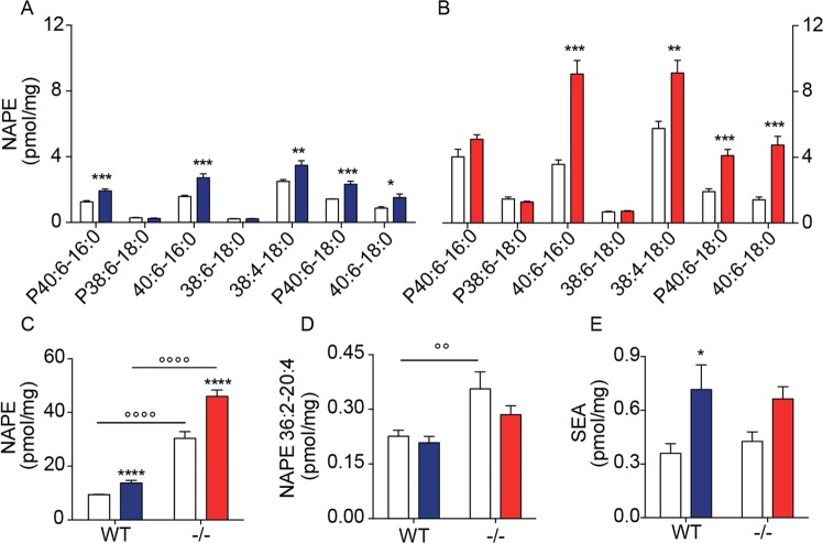
Effects of NAPE-PLD deletion on NAPE and FAE levels in mouse striatum. (A,B) Levels of individual NAPEs in dorsal striatum of (A) wild-type (WT) mice, and (B) NAPE-PLD−/− mice 48 h after intrastriatal 6-OHDA administration; open bars: control side of both WT and NAPE-PLD−/− mice; color-coded bars: lesioned side of WT (blue) or NAPE-PLD−/− (red) mice (n = 6–7). (C) Total NAPE levels in striatum of WT and NAPE-PLD−/− (−/−); open bars: control side for both WT and NAPE-PLD−/− mice; color-coded bars: lesioned side of WT (blue) or NAPE-PLD−/− (red) mice (n = 6–7). (D) Levels of anandamide precursor NAPE (36:2–20:4) in striatum of WT and NAPE-PLD−/− mice. Same color-coding as in (C) (n = 6–7). (E) SEA levels in striatum of WT and NAPE-PLD−/− mice. Same color-coding as in (C) (n = 6–7). *P < 0.05, **P < 0.01, ***P < 0.001, ****P < 0.0001 one-way ANOVA; °°°°P < 0.0001 two-way ANOVA, Bonferroni post hoc test.

