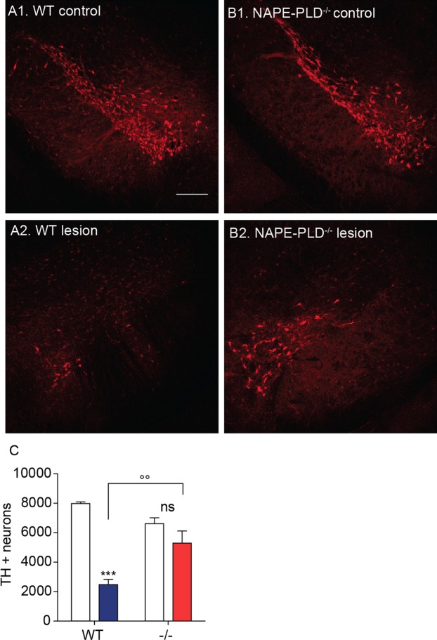
Figure 2.
Effects of NAPE-PLD deletion on 6-OHDA-induced neurotoxicity in mouse SN. (A,B) Immunofluorescence images for TH in tissue sections of SN pars compacta: (A1,B1) control and (A2,B2) lesioned (ipsilateral and contralateral, respectively, to the 6-OHDA injection site) of wild-type (A1–2) or NAPE-PLD−/− (B1-2) mice. Scale bar 200 µm. (C) Stereological count of TH+ neurons in SN pars compacta of WT and NAPE-PLD−/− mice. Open bars: control side of both WT and NAPE-PLD−/− mice; color-coded bars: lesioned side of WT (blue) or NAPE-PLD−/− (red) mice (n = 3–4). ***P < 0.001 compared to intact contralateral side; °°P < 0.01 compared to lesioned side of wild-type mice, two-way ANOVA, Bonferroni post hoc test.

