FIGURE 6.
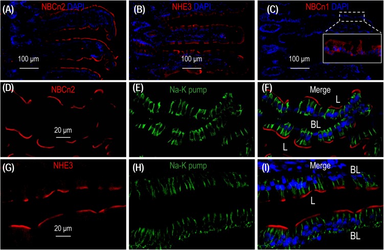
NBCn2 and NHE3 are expressed at the apical membrane of epithelium in small intestine. (A) Overview of staining with anti-MCDL (NBCn2) in the small intestine. (B) Overview of staining with anti-NHE3 in the small intestine. (C) Negative staining of anti-NBCn1 in the small intestine. In these experiments, the final concentrations of anti-MCDL, anti-NHE3, and anti-NBCn1 for immunofluorescence staining were 1.5 μg/ml (1:400 dilution). NBCn2 and NHE3 are mainly expressed in the villi of the small intestine. In panel C, no significant staining was observed for anti-NBCn1 when visualized under microscopy with parameters the same as those used for panel A and C. Inset in panel C shows that the non-specific background staining by anti-NBCn1, when visualized by a much higher exposure, is distributed throughout the cytosol of the epithelia. (D–F) High magnification view shows that NBCn2 is exclusively expressed at the apical membrane of small intestine epithelium. (G–I) High magnification view shows that NHE3 is exclusively expressed at the apical membrane of small intestine epithelium. In these experiments, the basolateral membrane is stained by α1 of Na+-K+ pump.

