Figure 3.
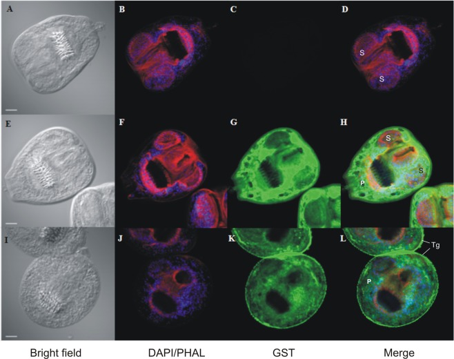
In toto immunolocalization of GST during PSCs oxidative stress response. PSCs were incubated in RPMI-10% FBS (C-PSCs) (A to H) or RPMI-10% FBS supplemented with 2.5 mM H2O2 (H-PSCs) (I to L). After PFA fixing, PSCs were incubated with anti-GST antibody (G and K) (GST) (green) and counterstained with DAPI-phalloidin (DAPI/PHAL) (see Materials and Methods section). Negative control consisted of omission of primary antibody (C). A, E, and I show the bright field and merge of the three channels in D, H and L. Scale bar = 20 μm. P: parenchyma: S: sucker: Tg: tegument.

