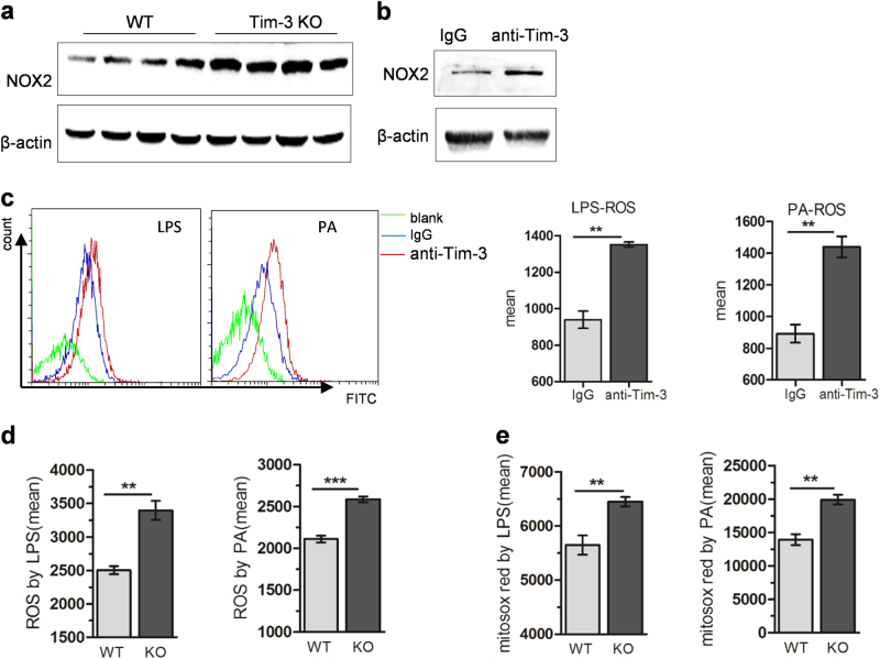
Fig. 4.
Tim-3 inhibited ROS generation in macrophages. a NOX2 expression was analyzed by western blot in liver tissues of WT and Tim-3 KO mice after MCD treatment. b Western blotting was performed to analyze NOX2 expression in BMDMs pretreated with anti-Tim-3-neutralizing antibody. BMDMs were pretreated with anti-Tim-3-neutralizing antibody following LPS or PA stimulation (c). Peritoneal macrophages prepared from Tim-3 KO or WT mice were treated with LPS or PA (d, e). ROS (c, d) and mitochondrial ROS (e) were assayed with DCFH-DA or mitosox red as described in the Materials and methods. **p < 0.01, ***p < 0.001

