Figure 1.
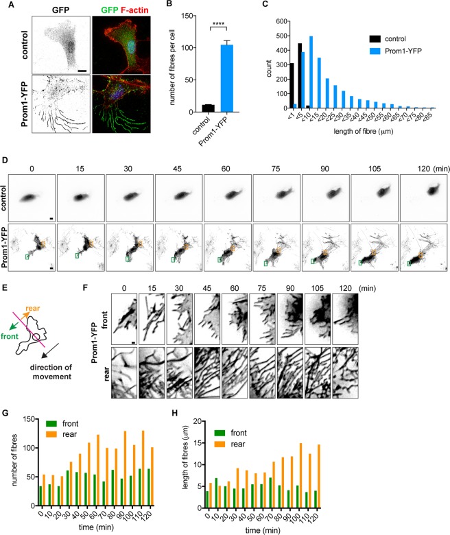
Prom1 induces cell membrane extensions. (A) Prom1-YFP induces fibre formation. Expression plasmids conveying control YFP or Prom1-YFP were transfected into the RPE1 cells and were harvested for 24 hours after the transfection. Cells were stained with GFP antibody (green) or phalloidin (red). (B,C) Quantitative data for the numbers (B) and lengths (C) of the fibres. In (B), 20 cells were analysed in each experiment, and the experiments were repeated four times. Data represent mean ± SE values of the four experiments. In (C), distribution of the fibre lengths measured on all the cells from four experiments are represented. (D) Live imaging analysis of the cells transfected with control (upper) or Prom1-expressing (lower) plasmids. Images were shown with 15 minute-intervals, starting at 24 hours after the Prom1 transfection. See also Supplementary Movie S1A and B. (E–H) The membrane extensions were mainly formed at the rear side against the direction of the migration. (E) The definition of the front and rear sides against the cell movement. (F) Focused images of the membrane extensions at the front (upper images) and at the rear (lower images) sides of the cell. (G,H) Quantitative data for the number (F) and length (G) of the fibres.

