Figure 5.
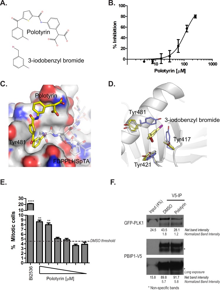
Targeting the Tyr pocket of the PLK1 PBD with a small-molecule inhibitor, Polotyrin. (A) The chemical structure of Polotyrin and 3-iodobenzyl bromide. (B) Polotyrin competitively inhibits the binding of a TAMRA- labelled-Glu-Thr-Phe(71)-Asp-Pro-Pro-Leu-His-pThr(78)-Ala-Ile-Tyr-Ala-Asp-Glu-acid phosphopeptide to the PLK1 PBD in Fluorescence Polarisation assay. (C) Crystal structure of PBD (surface rendering) with Polotyrin (stick model with yellow carbon atoms) shows how the compound binds in the Tyr-pocket that opens up on binding to long PBD substrates. Phosphopeptide FDPPLHSpTA from PBIP1 spanning the from Tyr pocket to the phospho-substrate binding groove is shown as transparent sticks for reference (PDB 3p37). (D) Complex of alpha-bromo-2-iodo-tolune (stick model with pale yellow carbons) bound to PBD with side chains of the aromatic residues aligning the Tyr pocket are shown as thin sticks (pale yellow). Tyr pocket lining residues from Polotyrin (yellow), FDPPLHSpTA (white) and LHSpTA (blue) complexes are also shown as well to illustrate structural changes in the pocket on binding to different ligands. (E) HeLa cells were treated with Polotyrin (at 1000, 800, 400, 200, 100 & 50 μM) and BI2536 (100 nM) for 24 h. Mitotic cells were scored as phospho-histone H3 positive cells and expressed as a percentage of the total number of Hoechst 33342-stained nuclei using a high-content screening platform as described earlier55. Each bar is a mean of three replicates ± S.D. and shows a dose-dependent increase in mitotic index upon treatment with Polotyrin; data from DMSO-treated cells is shown as a broken line. The data presented is representative of two independent experiments. Statistical analysis was done using Mann-Whitney two-tailed t-test; **p = 0.0029 to 0.0057; ****p < 0.0001. (F) HeLa GFP-PLK1Wt cells were transfected with PBIP1Wt-V5 and harvested 24 h later. The lysates were incubated with Polotyrin (2 mM) or DMSO. PBIP1Wt-V5 was pulled down using V5 antibody and co-immunoprecipitates were analysed by immunoblotting.

