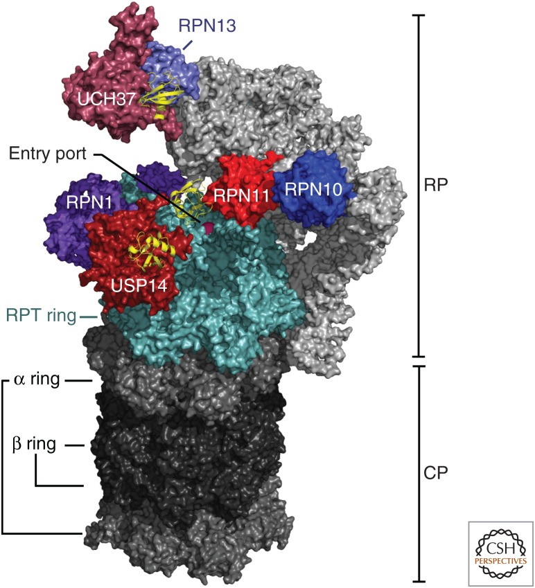
Figure 2.
Overview of deubiquitinating enzymes (DUBs) and Ub/Ubl receptors of the human proteasome. A cryo-electron microscopy (cryo-EM) density map of the human proteasome is represented with all of its known ubiquitin receptors (RPN1, RPN10, and RPN13 in purple tones) and DUBs (RPN11, USP14, and UCH37 in red tones). The lid subassembly of the RP is in light gray. The cryo-EM structure of the proteasome bound to USP14-ubiquitin-aldehyde was obtained from data presented in Huang et al. (2016b) (Protein Data Bank [PDB]: 5GJQ). The location of UCH37 and RPN13 were modeled as shown in de Poot et al. (2017) using data in density maps from Sahtoe et al. (2015) and Vander Linden et al. (2015) (PDB: 4UEL and 4WLQ). All six subunits of the RPT ring are shown in light blue and ubiquitin monomers bound to the DUBs are shown in yellow ribbon representations.

