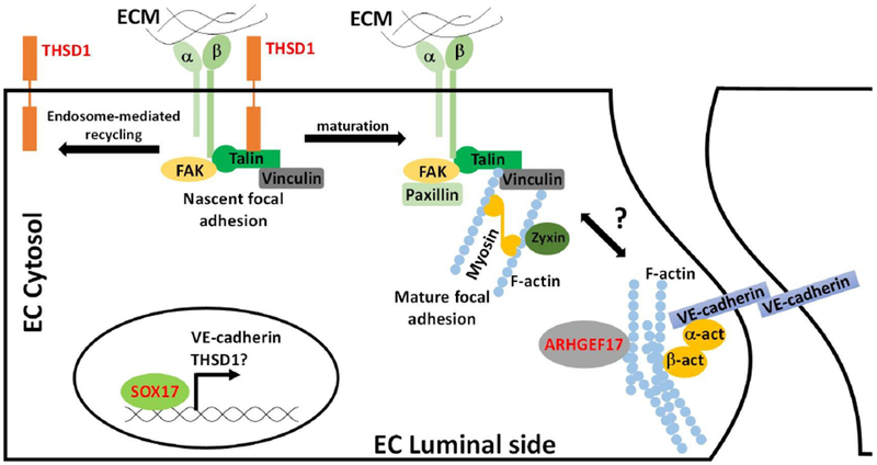
Figure 1. Schematic of IA genes in vascular endothelial cells.

Three IA genes including THSD1, SOX17, and ARHGEF17 are highlighted in red. THSD1 physically interacts with integrin complex through talin at nascent focal adhesion. When nascent focal adhesion matures, THSD1 leaves for next nascent focal adhesion site via endosome-mediated recycling process. Loss of THSD1 leads to defects in focal adhesion, a key determinant of the actin cytoskeleton, and modulator of several downstream pathways. Sox17 functions as a transcriptional factor and modulates VE-cadherin expression. Loss of VE-cadherin decreases cell-cell adhesion and increases permeability. ARHGEF17 is guanidine exchange factor that potentially regulates the remodeling of actin cytoskeleton.
