. 2019 Sep 18;19(5):e30. doi: 10.4110/in.2019.19.e30

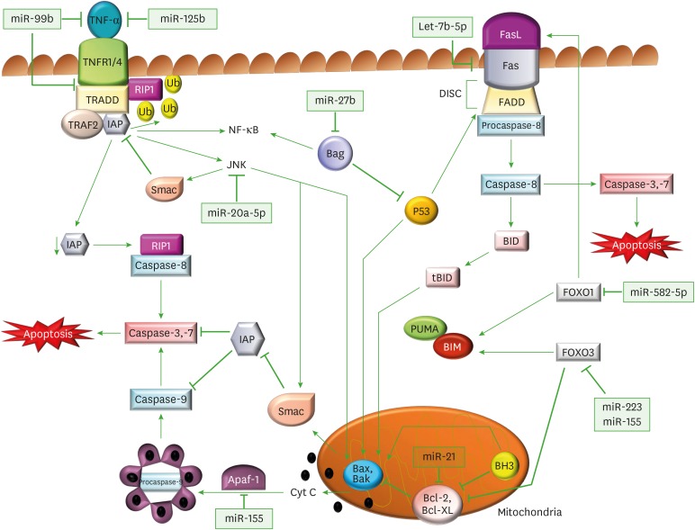
Figure 1. Regulation of apoptotic signaling pathways through miRs by M. tuberculosis. Diagrammatic representation of miRs and their target genes, which alter the canonical apoptotic pathways of immune cells, and assist in M. tuberculosis survival. In the extrinsic apoptotic pathway, TNFα binds to its receptors (TNFR1/4) and triggers its trimerization, which recruits TRADD, an adaptor molecule that allows binding of TRAF-2 and IAP to the receptor complex. RIP1, a kinase then binds TNFR1 through TRADD association. IAP proteins are responsible for RIP1 ubiquitination, and in their absence, RIP1 cannot be ubiquitinated. Non-ubiquitinated RIP1 can form a cytosolic complex with the caspase-8, leading to induction of apoptosis. The binding of TRAF-2 to the receptor complex stimulates the JNK pathway, which is a proapoptotic pathway. JNK can promote the release of Smac, from mitochondria, which inhibits TRAF2/IAP complex formation, and relieves the inhibition of caspase-8, thereby triggering caspase activation. JNK also participates in intrinsic apoptotic pathway, as after activation, JNK translocates to the mitochondria and phosphorylates BH3-only family of Bcl-2 proteins, which antagonize the anti-apoptotic activity of Bcl-2 or Bcl-Xl proteins. Further, JNK stimulates the release cyt C from mitochondrial inner membrane through BID-Bax dependent mechanism and promotes apoptosomes formation from released cyt C, caspase-9, and Apaf-1. The apoptosomes initiates caspase-9-dependent caspase cascade. In FasR pathway, a FasL binds to FasR and induces the recruitment of FADD followed by activation of procaspase-8, which further activates caspase-3 (the executioner) causing apoptosis. Caspase-8 also cleaves BID, a BH3 domain-containing protein of the BCL-2, which triggers intrinsic apoptosis and amplifies the signal from the extrinsic pathway. The expression of another member of BH3 only protein is increased by FOXO1 and FOXO3, which initiates the Bax/Bak-dependent apoptotic pathway.

Figure 1