. 2019 Sep 18;19(5):e30. doi: 10.4110/in.2019.19.e30

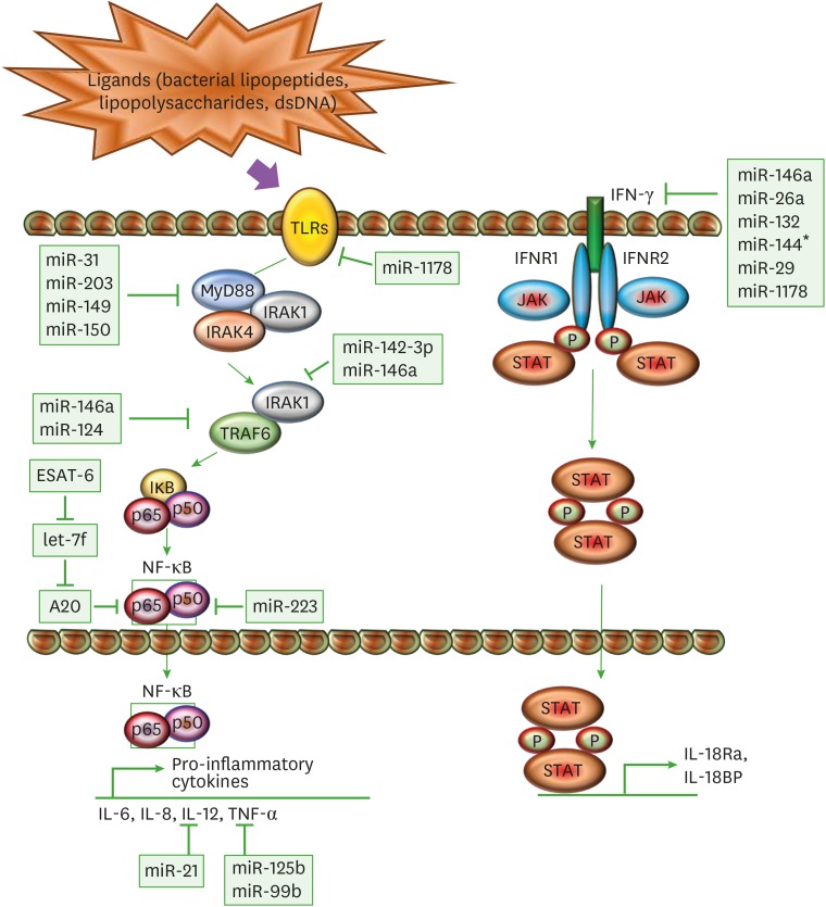
Figure 2. Immunomodulation of macrophages through miRs. A schematic diagram represents different miRs and their target genes, which allows M. tuberculosis to mitigate the immune response through modulation of TLR and IFN-γ pathway. The ligation of mycobacterial ligands to TLRs leads to the interaction of MyD88 with IRAK4 and IRAK1. IRAK4 auto-phosphorylates and activates IRAK1. This promotes the activation of TRAF6, which further activates TAK1. TAK1 gives rise to canonical IKK complex by phosphorylating IKKα and IKKβ, which phosphorylates components of the transcription factor NF-κB such as IκBα, p50, and p65. TAK1-mediated activation of NF-κB transcription factors drives the production of pro-inflammatory cytokines such as IL-12 and TNF-α. In another pathway, IFN-γ acts as a principal mediator of macrophage activation, which modulates pro-inflammatory cytokine production and induces production of anti-inflammatory molecules. Upon ligand binding, oligomerization and transphosphorylation of IFN-γ receptors (IFNR1 and IFNR2) activate JAK1 and JAK2, which creates a docking site for STAT1. STAT1 homodimerizes upon phosphorylation (P) in an antiparallel configuration, forming a complex γ-activated factor, which translocates to the nucleus and binds to the γ-activated site, located at the promoters of primary response genes, increasing their transcription, like IL-1Ra and IL-18BP. SOCS proteins negatively regulate the IFN-γ pathway by inhibiting JAKs and STAT1 phosphorylation.

Figure 2