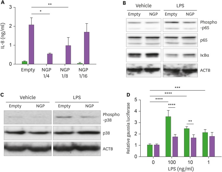
Figure 5. NGP blocks the activity of LPS. (A) THP-1 cells were treated by vehicle (black bars) or 100 ng/ml of LPS (blank bars) for 16 h in the mixture of NGP conditional medium and empty vector conditional medium in a ratio of 0, 1/4, 1/8, and 1/16. IL-8 was detected from the culture medium (quadruplet mean±SD). (B) THP-1 cells were treated by vehicle or 10 ng/ml of LPS in an empty vector or NGP conditional medium for 30 min. Phospho-NF-κB p65, NF-κB p65, and IκBα were detected by Western blotting. (C) THP-1 cells were treated by vehicle or 10 ng/ml of LPS in an empty vector or NGP conditional medium for 30 min. Phospho-p38 MAPK and p38 MAPK were detected by Western blotting. (D) The conditional medium of empty vector (black) or NGP (blank) was treated overnight. LPS was mixed in different concentrations and then treated to 293 cells transfected with Gaussia luciferase NF-κB construct. The luminescence of cells with NGP medium showed inhibited activity of the reporter (quadruplet mean±SD).
*p<0.05, **p<0.01, ***p<0.001, ****p<0.0001.

