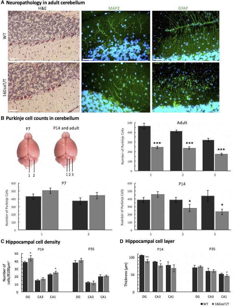
Figure 4.
Cerebellar and hippocampal pathology in 16GsoT/T mutants. (A) H&E staining showed that PCs are fewer in number and irregularly spaced in adult 16GsoT/T CB as compared to WT; MAP2 staining reveals that the 16GsoT/T PCs have sparse, irregularly branching dendritic trees, and GFAP staining highlights structural abnormalities of the Bergmann glia. (B) The number of PCs was quantified at different positions throughout the CB at different stages of development (P7, P14 and adult) in WT (black) and 16GsoT/T mutants (gray). Position 1 is at the midline, 1 and 2 each are 200 microns apart at P7, and 1, 2 and 3 each are 300 microns apart at P14, and 600 microns apart in adult. (C) Cell density in DG, CA3 and CA1 was quantified in WT (black) and 16GsoT/T (gray) at P14 and P35. 16GsoT/T has significantly increased cell number in DG and CA1 at P14. (D) Thickness of cell layer was measured in DG, CA3 and CA1 in WT (black) and 16GsoT/T (gray) at P14 and P35. n, WT = 16GsoT/T = 3 males at P7 and P14; WT = 16GsoT/T = 4-5 males in adult. ANOVA, *P < 0.05; **P < 0.01; ***P < 0.001. Scale bar = 50 μm.

