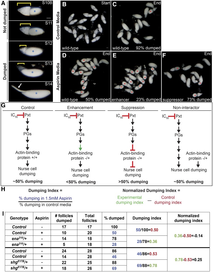
Figure 2.
In vitro egg maturation examples and screen rationale. A. Representative images of S10B-S14 Drosophila follicles taken using a stereo dissecting scope; anterior is at the left. Blue asterisks indicate the oocyte, yellow brackets mark the nurse cells, and the white arrow indicates the dorsal appendages in S14. In the IVEM assay, follicles that remain in S10B-11 are scored as having failed to dump, whereas follicles that have reached S12-14 are scored as having completed dumping. B-F. Representative images of in vitro maturing follicles at the start (B) or end of the experiment (C-F) in control media (B-C, vehicle) or aspirin media (D-F, IC50 aspirin). Follicles failing to dump are marked with red asterisks. G. Diagrams illustrating the rationale behind the pharmaco-genetic interaction screen. H. Formulas for calculating the dumping index and normalized dumping index (the colors match the data in the table in I). I. Table of the data and example calculations from two experiments – one for an enhancer (ena210) and one for a suppressor (shgE17B). In control media, the majority of wild-type follicles complete nurse cell dumping (C, 92%), while in aspirin media only 50% complete dumping (D, G). The dumping index for control follicles is expected to be around 0.5 (H, I). A genetic enhancer will result in <50% completing dumping in aspirin media (E [23%] and G), a dumping index of <0.5, and a negative normalized dumping index (I, ena210). A genetic suppressor will result in >50% completing dumping (F [73%] and G), a dumping index of >0.5, and a positive normalized dumping index (I, shgE17B). Reduced levels of an actin-binding protein that is not a downstream target of PG signaling during S10B would not be expected to modify follicle sensitivity to COX inhibition, resulting in 50% completing dumping (G), a dumping index of ∼0.5, and a normalized dumping index of ∼0. Scale bars = 50μm.

