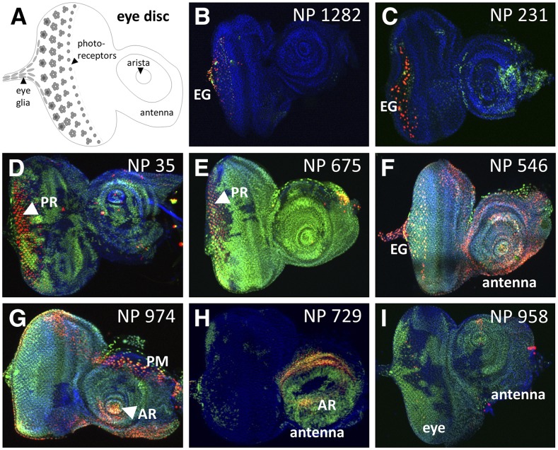
Figure 4.
Select GAL4-expressing lines with complex G-TRACE patterns in the eye disc. A) Schematic of the third instar larval eye disc showing the primary structures identified during screening. B-I) Fluorescence microscopy images showing various patterns of real-time GAL4 activity (RFP, red) and associated cell lineages (GFP, green) within the third instar larval eye disc. The corresponding NP line identifier is shown in the upper right corner of each image. For all images, DNA is shown in blue (DAPI staining). Eye glia (EG); photoreceptors (PR); arista (AR); peripodial membrane (PM).

