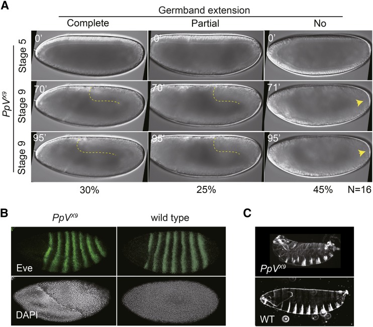
Figure 5.
PpV functions in pattern formation and germband extension. (A) Images from time-lapse movies of embryos from PpV germline clones with wide field optics. Dashed lines depict the posterior end of the germband. Arrow heads point to pole cells. Time in minutes (’). (B) Fixed embryos from wild type and PpV germline clones were stained for pair-rule protein Eve (green) and DNA (white). (C) Cuticles of embryos from wild type and PpV germline clones.

