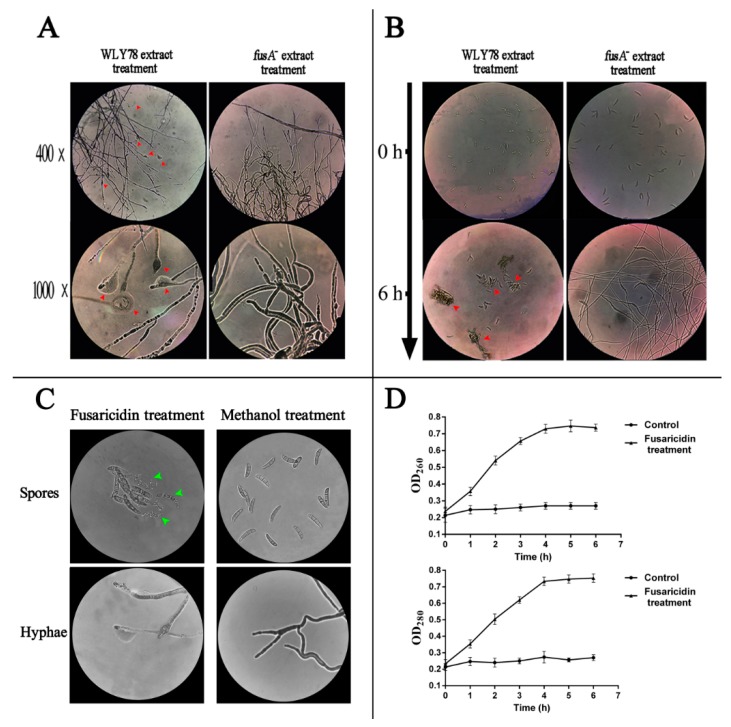
Figure 4.
400× and 1000× micrographs from an optical microscope for hyphae treated with WLY78 and the fusA− mutant extracts (A). The 100× micrograph from an optical microscope for the germination of spores treated with WLY78 and the fusA− mutant extracts for six hours (B). 1000× micrographs from an optical microscope for spores and hyphae treated with or without fusaricidin for two hours (C). Leakage levels of nucleic acid (OD260) and proteins (OD280) of F. oxysporum f. sp. cucumerium spore suspensions after treatment with or without purified fusaricidin were evaluated (D). The red arrows indicate the vesicle structures, the cytoplasm leakage, and spore aggregation. The green arrows indicate the spores bursting into pieces.

