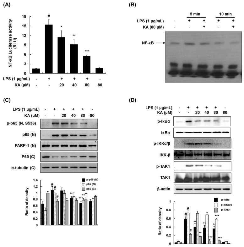
Figure 4.
Effects of KA on the nuclear factor-kappa B (NF-κB) signaling pathway in LPS-induced RAW 264.7 macrophages. (A) Cells were transiently transfected with pNF-κB-luc reporter; phRL-TK vector was used as an internal control. Cells were pretreated with KA (20, 40, or 80 μM) for 1 h and then stimulated with LPS (1 μg/mL) for 18 h. Luciferase activity levels were determined using the Promega Luciferase Assay System. (B) Nuclear extracts were prepared from cells stimulated with LPS at 5 and 10 min in the presence of KA (80 μM), and the DNA-binding activity of NF-κB was analyzed by EMSA. (C) Nuclear and cytosolic proteins were extracted from cells pretreated with KA (20, 40, or 80 μM) for 1 h and then stimulated with LPS for 5 min. The nuclear and cytosolic proteins were prepared, resolved by SDS-PAGE, and detected using specific p-p65 and p65 antibodies. Poly(ADP-ribose) polymerase-1 (PARP-1) and α-tubulin were used as internal controls for the nuclear and cytosolic fractions, respectively. (D) Cells were pretreated with KA (20, 40, or 80 μM) for 1 h and then stimulated with LPS for 5 min. Total cellular proteins were prepared, resolved by SDS-PAGE, and detected using specific p-IκBα, inhibitor of κB (IκBα), p-IKKα/β, IκB kinase (IKK-β), p-TAK1, and TGF-β-activated kinase 1 (TAK1) antibodies. β-actin was used as an internal control. Each experiment was performed three times, and similar results were obtained in each experiment. # P < 0.05 vs. the control group; * P < 0.05, ** P < 0.01, *** P < 0.001 vs. LPS-stimulated cells.

