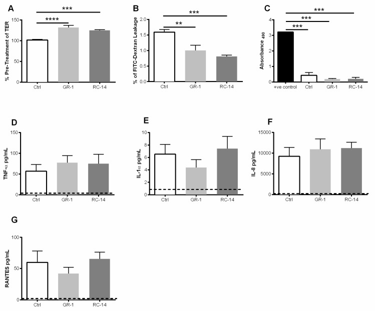
Figure 1.
Exposure to probiotic strains of lactobacilli improves barrier function of primary genital epithelial cells and does not alter viability or production of pro-inflammatory cytokines and chemokines. Confluent monolayers of GECs were treated for 2 h with probiotic strains L. rhamnosus GR-1 and L. reuteri RC-14 abbreviated GR-1 and RC-14 respectively and/or no bacteria (Ctrl). After treatment, non-adherent lactobacilli were removed, and GECs were monitored for a further 24 h. (A) TER was measured pre-treatment and at 24 h post-treatment with lactobacilli. TER is expressed as percent of pre-treatment TER. (B) Following treatment with bacteria, primary GECs were incubated with FITC-dextran dye on the apical surface, and FITC-dextran leakage in the basolateral compartment was measured after 24 h. Data are expressed as a percentage of FITC-dextran added to the apical compartment. (C) LDH release (Abs490) by GECs 24 h after treatment with GR-1, RC-14 or no bacteria, or from GEC cultures lysed to release total LDH (positive control). (D–G) 24 h post-treatment with lactobacilli, cell supernatants were collected and analyzed for the presence of TNF-α, IL-1α, IL-8, and RANTES. Dashed line indicates assay limit of detection. Data shown are representative of 6–9 separate experiments done on tissues taken from 6-9 different donors and shown as mean ± SEM. Each experiment had 3 replicate cultures for each experimental condition. Statistical significance is indicated as: **** p < 0.0001, *** p < 0.001, ** p < 0.01.

