Figure 3.
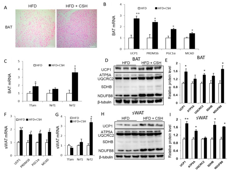
WECS activates BAT and induces browning of SWAT. Histology analysis showed that WECS treatment decreased the lipid droplets in the BAT(A). Scale bars, 100 μm. Besides, WECS treatment upregulated the expression of thermogenic genes and some mitochondrial genesis genes in BAT (B and C) and sWAT (F and G) in HFD mice. Besides, WECS upregulated the expression of UCP1 and some proteins related to oxidative phosphorylation in BAT (D) and sWAT (H). Relative protein levels of UCP1 and OXPHOS in BAT and sWAT (E and I). Bars represent the mean + SEM, n = 3–5. * p < 0.05, ** p < 0.01 compared with HFD control.

