Figure 5.
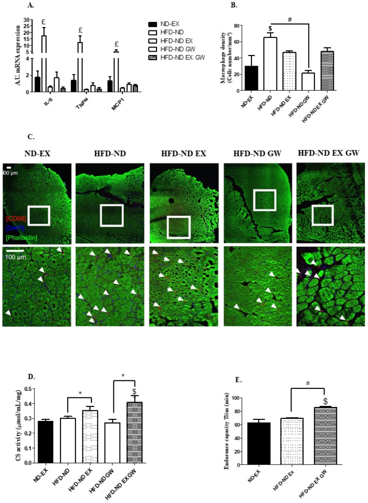
Inflammatory and metabolic states of skeletal muscle. (A) Relative mRNA levels of IL-6, TNF-α, and MCP-1. Data are expressed as arbitrary units of expression (A.U.) relatively to 36B4 in the vastus lateralis. (B,C) Macrophage infiltration by immunohistology staining with Alexa-Fluor488 (staining of actin filament) conjugated phalloidin, CD68 (staining of myeloid cells), and DAPI (staining of cell nucleus) in tibialis lateralis anterior. White borders show the selected area for magnification. White arrows represent infiltrated macrophages. Data are shown as macrophage density (cell number per mm²) for ND-EX (n = 3), HFD-ND (n = 6), HFD -ND-EX (n = 4), HFD-ND-GW (n = 6), and HFD-ND-EX-GW groups (n = 6). (D) Citrate synthase activity is expressed in µmol/mL/mg of vastus lateralis tissue. (E) Endurance performance is expressed as the time to exhaustion in minutes of treadmill running (Tlim) for exercise trained groups (ND-EX, HFD-ND-EX, HFD-ND-EX-GW). Data are expressed as mean ± SD. n = 6 per group. # p < 0.05 GW0742 effect; * p < 0.05 exercise training effect (2-way ANOVAs); $ p < 0.05 vs. ND-EX; £ p < 0.05 vs. all groups (one-way ANOVAs).

