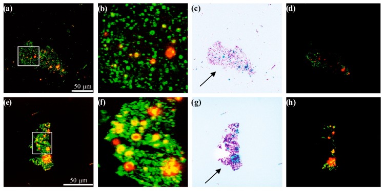
Figure 5.
Confocal microscopy images for Norit_1_6T (a–d) and Norit_2_6T (e–h) samples, (b) and (f) present enlarged details from confocal microscopy images shown in (a) and (e). (c) and (g) provide inverted-colour versions of the studied materials. (d) and (h) show the last recorded images after using the laser beam.

