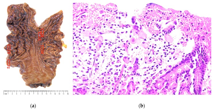
Figure 3.
(a) Prophylactic gastrectomy specimen with no visible macroscopic abnormalities. Complete mapping revealed five regions where microscopic foci of intramucosal signet ring cell carcinoma was found (red boxes). (b) Photomicrograph shows foci of signet-ring cell carcinoma, involving the gastric mucosa (stage IA disease) (Hematoxilin-eosin stain, 100X).

