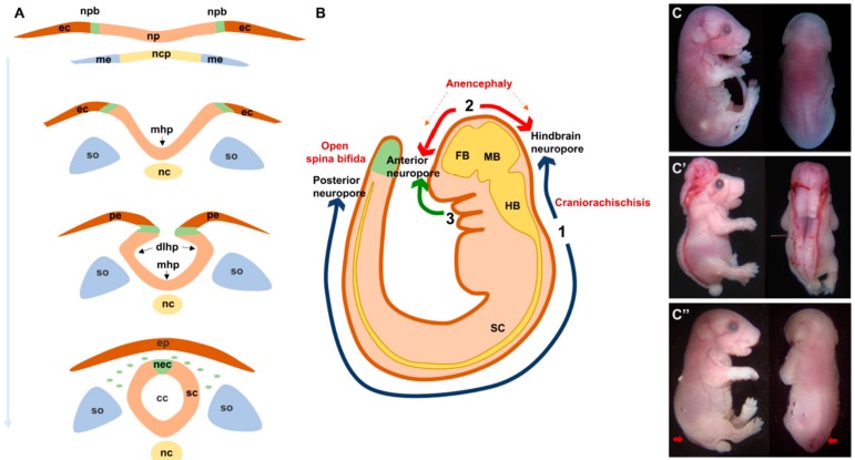
Figure 1.
Normal and abnormal neural tube formation. (A) Major steps of neural tube formation. The neural plate overlying the notochord elevates to form the neural folds around the midline, bends at the midline (mhp) and dorsolateral sites (dlhp) then fuses at the opposing tips of the neural folds and separates from the overlying epidermis. (B). Initiation sites of neural tube closure 1–3 in a mouse embryo. Defects in closure 1 and 2 lead to craniorachischisis and anencephaly respectively. Defects in closure at the caudal end of site 1 lead to open spina bifida. (C). Lateral and dorsal views of E18.5 embryos showing craniorachischisis (C′) and open spina bifida (C″, indicated by red arrows) as compared to wild type (C). cc, central canal; dlhp, dorsolateral hinge point; ec, ectoderm; ep, epidermis; fb, forebrain; hb, hindbrain; mb, midbrain; mhp, median hinge point; me, mesoderm; nc, notochord; ncp, notochordal plate; nec, neural crest; np, neural plate; npb, neural plate border; pe, presumptive epidermis; sc, spinal cord; so, somites.

