Figure 3.
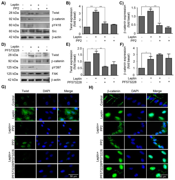
Src and FAK regulate the expression and subcellular localization of Twist and β-catenin. MCF10A cells were pre-treated with Src (PP2) or FAK (PF-573228) inhibitors, and subsequently with 400 ng/mL of leptin. (A) Representative western blots of Twist and β-catenin expression upon Src inhibition. Densitometric analyses of (B) Twist and (C) β-catenin expression upon Src inhibition. (D) Representative western blots of Twist and β-catenin expression upon FAK inhibition. Densitometric analyses of (E) Twist and (F) β-catenin expression upon FAK inhibition. β-actin was used as a loading control. The values represent the mean ± SD of three independent experiments and are expressed as changes with respect to the control (unstimulated cells). The asterisks indicate the comparison made with respect to the control. * p < 0.05 and *** p < 0.001 by one-way ANOVA (Newman–Keuls’s test). Representative images of epifluorescence microscopy using a 40× magnification. MCF10A cells were pre-treated with PP2 and PF-573228 and subsequently stimulated with leptin (400 ng/mL). The subcellular localization of (G) Twist and (H) β-catenin is shown in green, and in blue the DNA was counterstained with DAPI.

