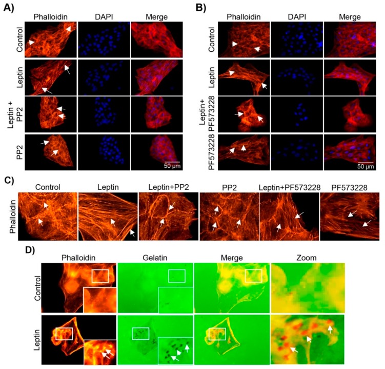
Figure 5.
Leptin stimulation leads to the re-arrangement of the actin cytoskeleton and formation of invadopodia. Representative images of MCF10A cells pre-treated or not with PP2 (A) or PF-573228 (B) for 30 min and subsequently with leptin (400 ng/mL) for 24 h. Actin filaments were detected with Phalloidin-TRITC (red) and DNA was counterstained with DAPI. Images were acquired using the 40× magnification. (C) Enlarged images of actin cytoskeleton of MCF10A cells grown in the presence or absence of leptin, and the Src and FAK inhibitors. White arrows indicate structures of actin. (D) Representative images of invadopodia formation assays. Actin puncta was detected with phalloidin (red) and Alexa 488-labeled gelatin (green) was used as a specific substrate for the membrane-bound MMP-14 located at the edge of invadopodia. Arrows indicate areas of gelatin degradation. Images were acquired using a 100× magnification.

