Figure 3.
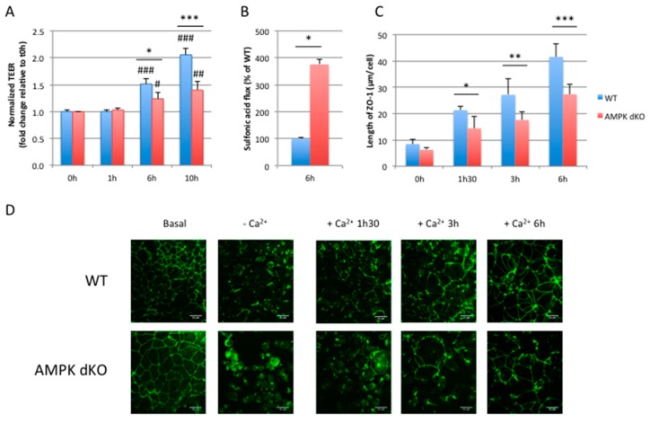
AMPK disruption delays barrier integrity recovery after calcium switch. (A) Time course of TEER development in WT and AMPK dKO Caco-2 cells subjected to a calcium switch. Cells grown on Transwell filters were incubated in calcium-free medium for 16 hours and switched to normal calcium medium. TEER was measured at the indicated time points after calcium switch and is given as fold change relative to the value in calcium-free medium at 0 h time point (0 h) (B) Paracellular permeability of 0.4 kDa FITC-sulfonic acid in WT and AMPK dKO Caco-2 monolayers subjected to calcium switch. Flux of FITC-sulfonic acid was measured at 6 h time point (6 h) and is given as the percentage of WT value. (C) Quantification of ZO-1 deposition at cell-cell junction after calcium switch at the indicated time points in WT and AMPK dKO Caco-2 cells. (D) Representative immunostaining of ZO-1 in WT and AMPK dKO Caco-2 cells at steady-state (basal), incubated in calcium-free medium for 16 hours (-Ca2+) or subjected to calcium switch (+Ca2+) for indicated time. Scale bar: 25 µm. Data represent means ± SD for three independent experiments (n = 3). * p < 0.05; ** p < 0.01; *** p < 0.005 versus AMPK dKO cells at the same time point. # p <0.05; ## p < 0.01; ### p < 0.005 versus 0 hour time point (0 h) for the same genotype.

