Figure 1.
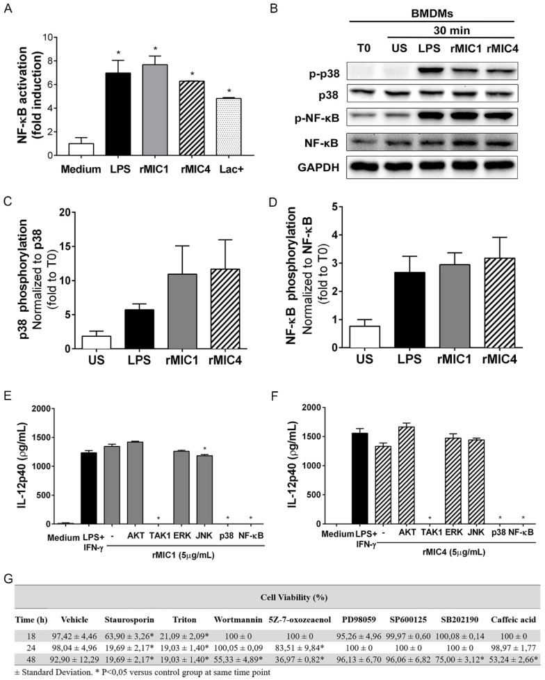
rMIC1 and rMIC4 induce IL-12 production by macrophages through activation of TGF-β activated kinase 1 (TAK1), p38, and nuclear factor-kappa B (NF-κB). (A) RAW264.7-luc murine macrophages were stimulated with the following preparations of Toxoplasma gondii microneme proteins: rMIC1 (5 µg/mL), rMIC4 (5 µg/mL), and the native Lac+ fraction (5 µg/mL), which contains MIC1 and MIC4. LPS (500 ng/mL) and medium were used as the positive and negative controls, respectively. Cells were lysed 4 h post-stimulation, and NF-κB activation was inferred from the luminescence measurements. Data are expressed as the mean ± SD of triplicate wells, and data from three independent experiments yielding similar results. (B) Total lysates of bone marrow-derived macrophages (BMDMs) was were 30 min after stimulation with rMIC1 (5 µg/mL), rMIC4 (5 µg/mL), or LPS (1 µg/mL). As controls, cells were also incubated with medium (i.e., unstimulated cells [US]) or sampled at time zero (i.e., T0), as indicated at the top of the panels. Immunoblotting was performed to assess total p38 and the p65 subunit of NF-κB, as well as their phosphorylated forms (p-p38 and p-NF-κB). Glyceraldehyde-3-phosphate dehydrogenase (GAPDH) was used as a loading control. (C,D) Densitometric analysis to quantify the p38 and NF-κB bands in the Western blot (panel B). (E,F) IL-12 concentrations in the supernatant of BMDM cultures that were pretreated with vehicle (-) or pharmacological inhibitors of AKT (Wortmannin, 100 nM), TAK1 (5Z-7-oxozeaenol, 100 nM), ERK (PD9805, 20 μM), JNK (SP600125, 20 μM), p38 (SB202190, 20 μM), or NF-κB (Caffeic acid, 15 µg/mL) for 3 h, and subsequently stimulated with (E) rMIC1 (5 µg/mL) or (F) rMIC4 (5 µg/mL) for 24 h as assessed by ELISA. LPS (1 µg/mL) and medium were used as the positive and negative controls, respectively. (G) Viability of BMDMs pretreated with pharmacological inhibitors of signaling molecules. Statistical analysis comparing the viability of cells (%) that were pretreated with each inhibitor and stimulated with rMIC1 or rMIC4 to that of cells stimulated with rMICs but not pretreated with pharmacological inhibitors (vehicle). Staurosporine (2 µM) and Triton X-100 (1% v/v) were used to induce in vitro apoptosis. The data are from two independent experiments yielding similar results. (*) p < 0.05 by one-way ANOVA, followed by Bonferroni’s post-test.

