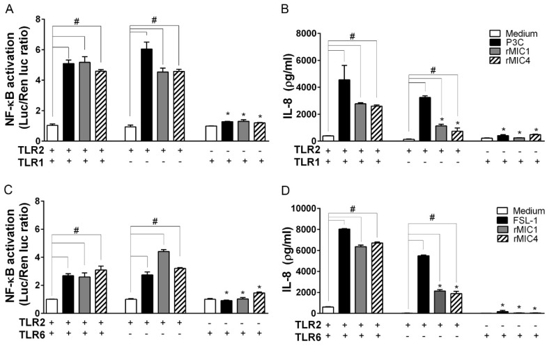
Figure 3.
TLR2 is critical for cell activation stimulated by microneme proteins. HEK293T cells were transfected with TLR2/1, only TLR1, or only TLR2 (A,B) or with TLR2/6, only TLR6, or only TLR2 (C,D). The cells were also co-transfected with the co-receptors CD14 and CD36, the NF-κB-dependent luciferase reporter construct (pELAM-firefly luciferase), and a Renilla luciferase reporter construct (an internal control). The total amount of DNA in each transfection was kept constant by adding empty expression vector. After 48 h of transfection, the cells were stimulated with rMIC1 (50 nM) or rMIC4 (50 nM). The positive controls for cell stimulus were Pam3CSK4 (P3C, 1 nM) for TLR2/1-transfected cells and FSL-1 (1 nM) for TLR2/6-transfected cells. Medium was used as the negative control. Cells were lysed 4 h after stimulation, and NF-κB activation was inferred from the luminescence measurements. Cell supernatants were harvested 24 h after stimulation, and the IL-8 concentration was assessed by ELISA. (+) Cells expressing the receptor and (-) cells without expression of receptor. In the statistical analysis, the responses of (*) cells lacking a receptor were compared to those of cells expressing heterodimers (TLR2/1 or TLR2/6). In addition, in cells expressing combinations of receptors or a single receptor, statistical comparisons (#) of responses the elicited by rMIC proteins versus the responses in the negative control (medium) were also performed. The data are from three independent experiments yielding similar results. (*) or (#) p < 0.05 by one-way ANOVA followed by Bonferroni’s test.

