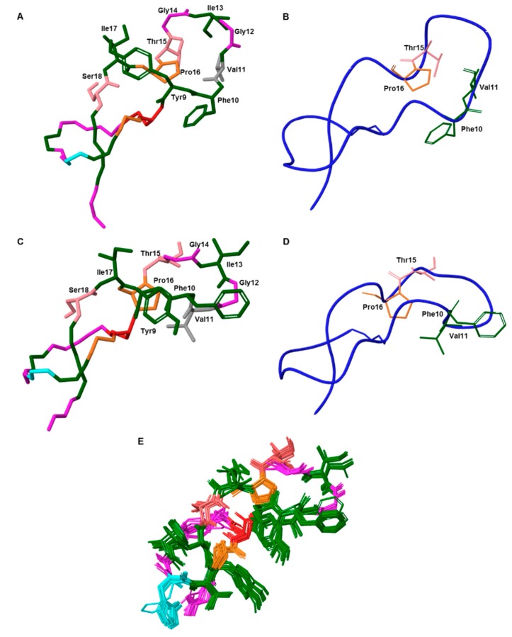
Figure 6.
(A) and (C) 3D structure of the native MccJ25 (PDB code 1PP5) and NMR-derived lowest-energy structure of the new compound, respectively. Only the side chains of the loop are shown. (B) and (D) Ribbon representation of the native peptide and new compound, respectively. The four residues involved in the β-sheet located in the loop are shown. (E) Ensemble of the 20-lowest energy structures derived from the NMR restraints of the new compound. The maximum RMSD value between structures 1–20 is 0.6 Å. Aromatic and hydrophobic residues are shown in green, negatively charged residue (Glu8) in red, positively charged residues (His5) in blue, polar residues in light pink and non-polar residues in pink (Gly) and (Pro). In (A) and (C) Val11 residue is shown in grey.

