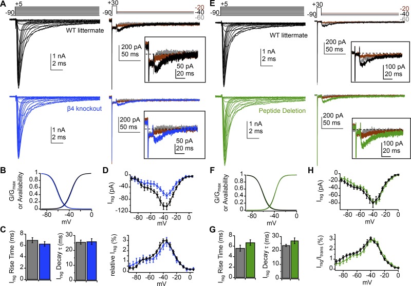
Figure 2.
Retention of normal transient and resurgent currents in NaVβ4−/− and peptide deletion Purkinje cells. (A) Transient (left) and resurgent (right) Na current from a WT (black) and NaVβ4−/− (blue) isolated Purkinje cell (50 mM extracellular Na, 5 nM TTX). Inset, resurgent component at higher gain. (B) Activation and inactivation curves with mean parameters for WT (n = 6) and NaVβ4−/− (n = 8) Purkinje cells (values in text). (C) Rise time (left) and decay τ (right) of resurgent current at −30 mV for WT and NaVβ4−/− (values in text). (D) Peak absolute resurgent current versus voltage (top) and relative resurgent current versus voltage (bottom) for WT and NaVβ4−/−. (E–H) Same as A–D but for cells from peptide deletion mice (green, n = 9) and WT littermates (black, n = 9). Resurgent current at −30 mV, −70.3 pA (WT), −92.1 pA (mutant); resurgent to transient ratios, 3.4% (WT), 3.2% (mutant). Data in C, D, G, and H are mean ± SEM.

