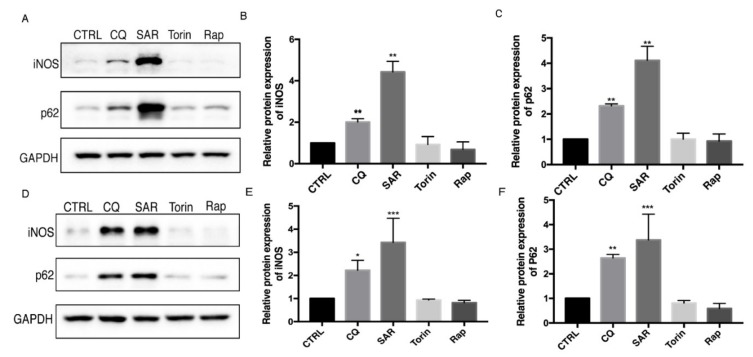
Figure 1.
Autophagy inhibition lead to iNOS and p62 accumulation in Raw 264.7 and bone marrow-derived macrophages (BMDM). (A,D) Raw 264.7 cells (A–C) and BMDMs (E–F) were incubated with CQ (30 µM), SAR (1 µM), Torin (1 µM), Rap (1 µM) for 24h. iNOS, p62 levels were detected by western blotting. CTRL is blank control group with DMSO treatment. (B,E) Western blot analysis of iNOS in Raw 264.7 cells and BMDM cells under basal condition. (C,F) Western blot analysis of p62 in Raw 264.7 cells and BMDM cells under basal condition. (B–F) Values were analyzed from three individual experiments by GraphPad Prism, each experiment conducted three times. * p < 0.05, ** p < 0.01, *** p < 0.001. Error bars (mean ± SEM). One-way ANOVA with Turkeys as post hoc tests.

