Figure 5.
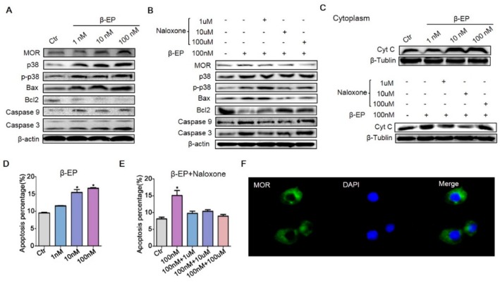
β-EP exposure induces apoptosis of TM3 cells. (A) Elevated expression levels of MOR, p38 MAPK, Bax, Bcl-2, caspase 9, and caspase 3 in TM3 cells after β-EP treatment as evidenced by western blotting. (B) Reversal of these effects by naloxone. (C) Elevated Cyt C after β-EP treatment and reversal by naloxone. (D) Elevated apoptosis and protection by naloxone as measured by flow cytometry. (E) Elevated apoptosis after β-EP treatment and reversal by naloxone as measured by flow cytometry. (F) Immunofluorescent staining of MOR (green) and DAPI (blue) in TM3 cells.

