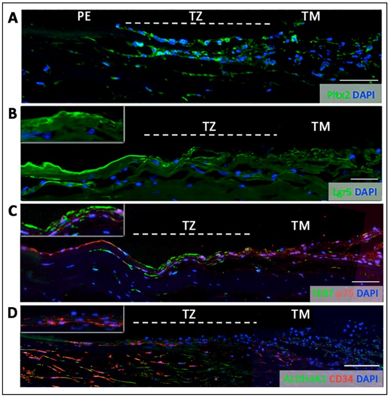
Figure 4.
Immunostaining on transverse sections showing the expression of (A) nuclear Pitx2 across the entire transition zone (TZ). The stem cell markers, (B) Lgr5 on cell surface, (C) telomerase (TERT) and (D) CD34 were more expressed in the inner TZ. The positive cells were also located in the stroma immediately beneath the TZ surface (insets). (D) Quiescent stromal keratocytes co-expressed CD34 and aldehyde dehydrogenase 3A1 (ALDH3A1, a keratocyte marker). White dotted lines delineate TZ location between peripheral endothelium (PE) and trabecular meshwork (TM). Scale bars: 50 μm.

