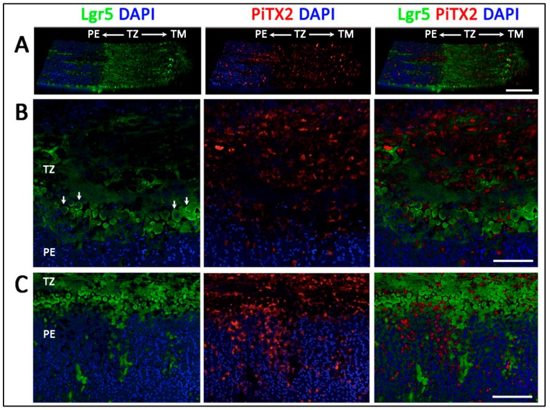
Figure 5.
Immunolocalization of Lgr5 and Pitx2 in inner transition zone (TZ) and peripheral endothelium (PE) regions. (A) A horizontal view tilted at 30° showed Lgr5 predominantly localized in inner TZ while Pitx2 staining had a pan-TZ pattern. (B) Lgr5 and Pitx2 expression in inner TZ region, next to PE. (C) Lgr5 positive cell clusters extended from inner TZ into PE. Scale bars: 100 μm.

